

Displaced intra-articular fractures of the hallux require reductionįractures of the phalanx are the most common type of foot fractures in the pediatric population.Nail-bed injuries involving the germinal matrix should be repaired.Open fractures require irrigation & debridement.Often not treated by orthopedic surgeons.One of the most common foot fractures in children.Plate removal should not be done routinely, and, if needed, it should not be done earlier than 18 months after surgery because of the risk of refracture.Study Guide Foot Fractures - Phalanx Key Points: ComplicationsĬomplications of surgery for Galeazzi fractures are similar to those for other forearm fractures-nonunion, delayed union, malunion, nerve injuries and infection. These can be treated, first, with distal radius articular restoration, and then, with the standard treatment of the Galeazzi fracture. Transitional InjuriesĬertain high-energy metadiaphyseal injuries are essentially a combination of distal radius fracture and a Galeazzi fracture. However, closed reduction is often successful in children. These injuries, termed Galeazzi equivalents, are normally treated the same way as a Galeazzi fracture. In adults, a fracture of the radial shaft can be associated with an additional fracture of the distal ulna. In children, a fracture of the radial shaft is sometimes associated with the separation of the distal ulnar epiphysis without disruption of the DRUJ. Alternative fixation, such as Kirschner wires and intramedullary pins, are not rigid enough to resist deforming forces and are unable to control rotation and allow shortening of the radius. DRUJ stability should be carefully assessed intraoperatively and addressed accordingly.ĭeforming forces (e.g., from the brachioradialis, pronator quadratus and the weight of the hand) make it difficult to control these fractures in a cast, hence there is a high percentage of failure with non-operative management.
GALEAZZI FRACTURE POSNA HOW TO
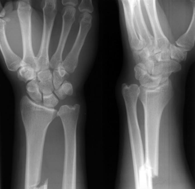
Plate and screw fixation is the preferred method, and the review describes the technique in detail, including how to address comminution. Treatment of AdultsĪdults with a Galeazzi fracture require open surgery for anatomic and rigid fixation of the radius shaft and stabilization of the DRUJ. Computed tomography (CT) or a radiograph of the contralateral wrist can be useful for assessing the DRUJ. In some cases, injury to the DRUJ is purely ligamentous. More than 5 mm of shortening of the radius relative to the ulna.Dislocation of the radius relative to the ulna (on a true lateral view).Widening of the DRUJ space (on a true AP view).Fracture at the base of the ulnar styloid.Neurovascular damage is rare.Įssential imaging includes good anteroposterior (AP) and lateral radiographs of the forearm, as well as orthogonal views of the wrist and elbow. Galeazzi patients usually present with swelling and deformity of the forearm. Mudgal, MD, in the Hand and Arm Center at Massachusetts General Hospital, reviewed the evaluation and treatment of Galeazzi fractures, which are sometimes called Piedmont fractures, reverse Monteggia fractures or Darrach–Hughston–Milch fractures. In Hand Clinics, orthopedic surgeons Rohit Garg, MD, MBBS, and Chaitanya S. These injuries usually occur as a result of high-energy trauma (e.g., motor vehicle accident, sports injury or a fall from a height) or a fall on an outstretched and pronated hand. Error: Please enter a valid email address.įractures of the radius shaft associated with a dislocation of the distal radioulnar joint (DRUJ) are called Galeazzi fractures.


 0 kommentar(er)
